Migraines are vascular headaches. An abnormal stimuli in the trigeminal nerve trigger the swelling of blood vessels carrying nerves. Abnormal stimuli in the trigeminal nerve cause it to release chemicals that trigger the expansion of blood vessels. The causes excruciating headaches, neck pain and swelling on the face.
The headache occurs when trigeminal nerve is stimulated. The expressed form of gene expressions of intrinsically photosensitive retinal ganglion cells mediate the pain which are present in the pathways of the trigeminal part of the bran or the 5th cranial nerve. This nerve sends impulses (including pain impulses) from the eyes, scalp, forehead, upper eyelids, mouth, and jaw to the brain.
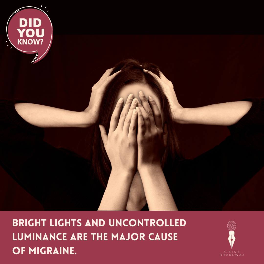
Bright lights and uncontrolled luminance are the major cause of migraine. Light can modify trigeminal activity without involving the central visual system. Photophobia is very common in state of migraine during pre and post migraine attacks, migraineurs are very sensitive to light.
Light and other visual stimuli also can trigger migraine attacks: for example, flickering or pulsing lights, repetitive patterns, glare, bright lights, computer screens, TV, and movies. Fluorescent light contains invisible pulsing, which is likely why so many report it as a migraine trigger.
The photoreceptors in the retina that are responsible for forming images typically comprise cones and rods. The visible and non-visible form of light inputs to the optical through the retina are encoded and projected to the primary visual cortex through RODS and CONES. This pathway is called the image forming pathway.
In non-imaging pathway the intrinsically photosensitive RGCs (ipRGCs) are found to be responsible of mediating the values of perceived brightness through the surge of eV of the retinal nerve which is expressed by melanopsin (their maximum spectral sensitivity is approximately 480 nm). In addition, ipRGCs receive inputs from cones and rods. Since it is already established and known that the light signals received by the melanopsin-expressing RGCs are projected to the suprachiasmatic nucleus (SCN), which entrains circadian rhythms, and to the olivary pretectal nucleus (OPN), which controls the pupillary light reflex, thus there are many evidence based researches that the projection to the superior colliculus and the contribution to migraine-associated photophobia suggests that melanopsin signals are involved in other non-image-forming visual effects as well. Therefore, according to various research establishments, even if a visual stimulus with an equi-luminance is presented, the pupil diameter may change when the melanopsin stimulus intensities are different.
On the basis on the experimental results, researchers formulated brightness perception using the stimuli on melanopsin and cones, and the pupil diameter as explanatory variables. These results showed that the melanopsin is not a minor contributor in brightness perception but, rather present as a critical factor for the formulation.
The perceived brightness differs, even with the same luminance or the same retinal illuminance, if the stimulus intensity on melanopsin is different over a luminance range. Statistical tests were conducted to investigate the difference in brightness perception between stimuli comparing different M/P ratios [Melanopic / Photopic] for same recommended luminance values cd/m2. This clearly demonstrated that melanopsins are involved in brightness perception. (Brown et al. revealed the relationship between the test radiance at equal brightness and the melanopic excitation of the reference stimulus.)
In addition, cones are easily adapted by light. Therefore, absolute brightness information relating to the light environment is not acquired by cones. The studies have revealed the facts that the relation between the visual stimulus intensity for melanopsin and the perceived brightness is linear, which suggests that the absolute brightness information may be coded under relatively limited light adaptation of melanopsin.
Thus, melanopsin appear to determine the perceived brightness level, and cone responses are added to it. In the non-image forming pathway, circadian rhythm photo-entrainment is performed by projecting from ipRGCs to the SCN. In this projection pathway, absolute intensity information regarding the light environment may be also necessary for controlling biological functions.
Compared with elderly people, young people have less influence of age-related changes of crystalline lens and vitreous body dysfunction. If brightness perception targeting a wide range of age groups is formulated, correction of spectral power distributions would be required.
“Photophobia is triggered by melanopsin and cone luminance inputs to the cortex via the (RTC) retino-thalamo-cortical pathway. Researchers have found that the adopted artificial lighting strategies incorporating luminaires or the light sources with low melanopsin excitation and photopic luminance to limit the lighting conditions leading to photophobia.”(Scientific reports -Google scholar)
![Migraines are vascular headaches. An abnormal stimuli in the trigeminal nerve trigger the swelling of blood vessels carrying nerves. Abnormal stimuli in the trigeminal nerve cause it to release chemicals that trigger the expansion of blood vessels. The causes excruciating headaches, neck pain and swelling on the face. The headache occurs when trigeminal nerve is stimulated. The expressed form of gene expressions of intrinsically photosensitive retinal ganglion cells mediate the pain which are present in the pathways of the trigeminal part of the bran or the 5th cranial nerve. This nerve sends impulses (including pain impulses) from the eyes, scalp, forehead, upper eyelids, mouth, and jaw to the brain. Bright lights and uncontrolled luminance are the major cause of migraine. Light can modify trigeminal activity without involving the central visual system. Photophobia is very common in state of migraine during pre and post migraine attacks, migrainuers are very sensitive to light. Light and other visual stimuli also can trigger migraine attacks: for example, flickering or pulsing lights, repetitive patterns, glare, bright lights, computer screens, TV, and movies. Fluorescent light contains invisible pulsing, which is likely why so many report it as a migraine trigger. The photoreceptors in the retina that are responsible for forming images typically comprise cones and rods. The visible and non-visible form of light inputs to the optical through the retina are encoded and projected to the primary visual cortex through RODS and CONES. This pathway is called the image forming pathway. In non-imaging pathway the intrinsically photosensitive RGCs (ipRGCs) are found to be responsible of mediating the values of perceived brightness through the surge of eV of the retinal nerve which is expressed by melanopsin (their maximum spectral sensitivity is approximately 480 nm). In addition, ipRGCs receive inputs from cones and rods. Since it is already established and known that the light signals received by the melanopsin-expressing RGCs are projected to the suprachiasmatic nucleus (SCN), which entrains circadian rhythms, and to the olivary pretectal nucleus (OPN), which controls the pupillary light reflex, thus there are many evidence based researches that the projection to the superior colliculus and the contribution to migraine-associated photophobia suggests that melanopsin signals are involved in other non-image-forming visual effects as well. Therefore, according to various research establishments, even if a visual stimulus with an equi-luminance is presented, the pupil diameter may change when the melanopsin stimulus intensities are different. On the basis on the experimental results, researchers formulated brightness perception using the stimuli on melanopsin and cones, and the pupil diameter as explanatory variables. These results showed that the melanopsin is not a minor contributor in brightness perception but, rather present as a critical factor for the formulation. The perceived brightness differs, even with the same luminance or the same retinal illuminance, if the stimulus intensity on melanopsin is different over a luminance range. Statistical tests were conducted to investigate the difference in brightness perception between stimuli comparing different M/P ratios [Melanopic / Photopic] for same recommended luminance values cd/m2. This clearly demonstrated that melanopsins are involved in brightness perception. (Brown et al. revealed the relationship between the test radiance at equal brightness and the melanopic excitation of the reference stimulus.) In addition, cones are easily adapted by light. Therefore, absolute brightness information relating to the light environment is not acquired by cones. The studies have revealed the facts that the relation between the visual stimulus intensity for melanopsin and the perceived brightness is linear, which suggests that the absolute brightness information may be coded under relatively limited light adaptation of melanopsin. Thus, melanopsin appear to determine the perceived brightness level, and cone responses are added to it. In the non-image forming pathway, circadian rhythm photo-entrainment is performed by projecting from ipRGCs to the SCN. In this projection pathway, absolute intensity information regarding the light environment may be also necessary for controlling biological functions. Compared with elderly people, young people have less influence of age-related changes of crystalline lens and vitreous body dysfunction. If brightness perception targeting a wide range of age groups is formulated, correction of spectral power distributions would be required. “Photophobia is triggered by melanopsin and cone luminance inputs to the cortex via the (RTC) retino-thalamo-cortical pathway. Researchers have found that the adopted artificial lighting strategies incorporating luminaires or the light sources with low melanopsin excitation and photopic luminance to limit the lighting conditions leading to photophobia.”(Scientific reports -Google scholar) It’s been 200 years the artificial light was experienced by very few population of the world and it’s approximately 100 years that people living in urban, suburban and few of the villages have experienced the artificial form of light. Even in these 100 years the form of light changed from Incandescent, IRC, mercury, metal halide and now LEDs. Daylight is the unpolluted form of healthy lighting since our eyes have had several million times longer to optimize to daylight than to LEDs. It is therefore reasonable to proclaim that human eyes, and behaviour are not yet optimized to LEDs. (As it’s only been one decade we are being used to it subjected with an abused form of it) With the change in the DNA of light source from heat energy to PN junction sources we need to consider visual and non-visual effects of light on people. However, there many guidelines and regulations from IES (Illuminating engineering Society), CIE, ANSI, IWBI, USGBC with revised lighting standards that address visual and non-visual aspects like visual comfort and circadian drive but now there is a huge gap and conflicts amongst the understanding of physiological aspects and working on those standards only and completely ignoring the flip side of adhering to those advantages. In the name of circadian drive we are forgetting the circadian alignment and synchronisation from one physiological state to another, from one environment to another and most importantly the nausea, headaches, change in the texture of skin and mediating migraines because of achieving the defined Equivalent melanopic lux (ma a time creating the bright ceilings or bright walls etc.) In view of the new standards to help the circadian drive and not the alignment is creating physiological imbalances w.r.t. the immune system, regeneration of the degenerated molecular cells, and neuronal activities. Apart from this, the melanopsin which are the cause of mediating the migraine if the these set of ipRGCs are subjected to high luminance or bright lights (even for blind persons) are totally ignored in the name of EML or the horizontal illuminance of 500 lx for workplaces etc. Similarly a higher illuminance value with the colder wavelengths, which is to be used e.g., in rooms with elderly persons with lower eyesight and to help them not entering in the zone of depression have to be prudently designed with an appropriate form of the luminaires and their directions or else it will definitely triggers the painful headaches and migraines in them making them more sick. The adherence to the lighting standards like illuminance levels, luminance ratios, glare controls, UGR, EML and circadian stimulus (CS) laid by these organisation is a must. However, mostly it is being misunderstood, misused and misguided by many of us. This is because either we are unaware or lack in the physiology associated to light as a zeitgeber. “That’s why I advocate that, lighting fraternity is no less than the medical fraternity”](https://yellout.in/girish/wp-content/uploads/2021/10/Theralicht-presentation-part-1-1024x576.png)
It’s been 200 years the artificial light was experienced by very few population of the world and it’s approximately 100 years that people living in urban, suburban and few of the villages have experienced the artificial form of light. Even in these 100 years the form of light changed from Incandescent, IRC, mercury, metal halide and now LEDs.
Daylight is the unpolluted form of healthy lighting since our eyes have had several million times longer to optimize to daylight than to LEDs. It is therefore reasonable to proclaim that human eyes, and behaviour are not yet optimized to LEDs. (As it’s only been one decade we are being used to it subjected with an abused form of it)
With the change in the DNA of light source from heat energy to PN junction sources we need to consider visual and non-visual effects of light on people. However, there many guidelines and regulations from IES (Illuminating engineering Society), CIE, ANSI, IWBI, USGBC with revised lighting standards that address visual and non-visual aspects like visual comfort and circadian drive but now there is a huge gap and conflicts amongst the understanding of physiological aspects and working on those standards only and completely ignoring the flip side of adhering to those advantages.
In the name of circadian drive we are forgetting the circadian alignment and synchronisation from one physiological state to another, from one environment to another and most importantly the nausea, headaches, change in the texture of skin and mediating migraines because of achieving the defined Equivalent melanopic lux (ma a time creating the bright ceilings or bright walls etc.)
In view of the new standards to help the circadian drive and not the alignment is creating physiological imbalances w.r.t. the immune system, regeneration of the degenerated molecular cells, and neuronal activities. Apart from this, the melanopsin which are the cause of mediating the migraine if the these set of ipRGCs are subjected to high luminance or bright lights (even for blind persons) are totally ignored in the name of EML or the horizontal illuminance of 500 lx for workplaces etc.
Similarly a higher illuminance value with the colder wavelengths, which is to be used e.g., in rooms with elderly persons with lower eyesight and to help them not entering in the zone of depression have to be prudently designed with an appropriate form of the luminaires and their directions or else it will definitely triggers the painful headaches and migraines in them making them more sick.
The adherence to the lighting standards like illuminance levels, luminance ratios, glare controls, UGR, EML and circadian stimulus (CS) laid by these organisation is a must. However, mostly it is being misunderstood, misused and misguided by many of us. This is because either we are unaware or lack in the physiology associated to light as a zeitgeber.
“That’s why I advocate that, lighting fraternity is no less than the medical fraternity”

